The Glute Muscles
Anatomy, Structure, and Biological Function
The gluteal muscles form a complex anatomical system responsible for hip movement, pelvic positioning, and force transfer between the upper and lower body. Although commonly discussed as a single group, each glute muscle has distinct structural characteristics and biological roles.
This guide serves as a reference overview of glute anatomy, detailing where each muscle is located, how it is structured, and what it is designed to do, independent of specific exercises or training methods.
What This Guide Covers
- The anatomical layout of the gluteal muscles
- Origin and insertion points
- Muscle fiber orientation and structure
- Primary and secondary biological actions
- The role of surrounding stabilizing muscles
Overview of the Gluteal Muscle Group

The Glutes as an Anatomical System
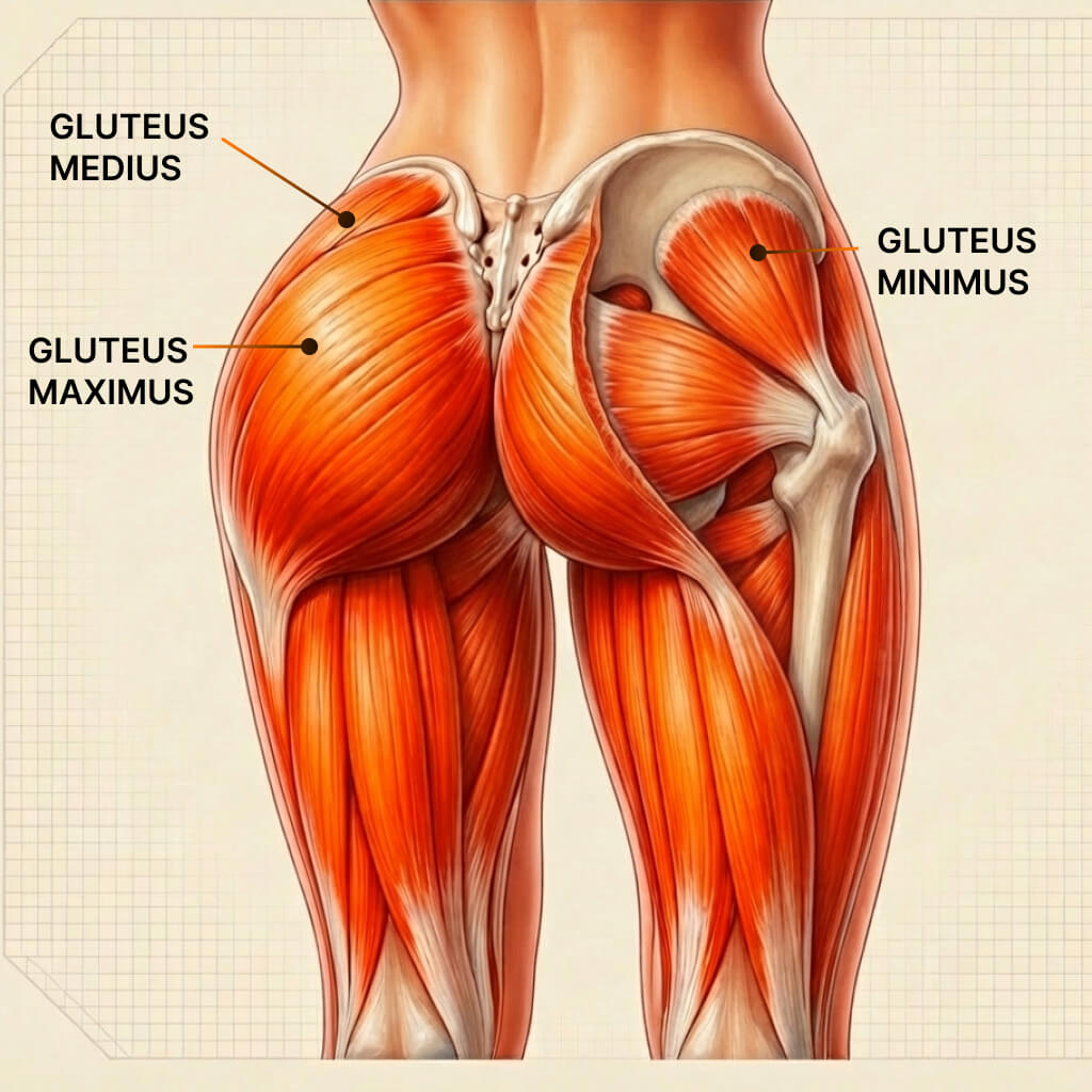
The gluteal region consists of three primary muscles
- Gluteus maximus
- Gluteus medius
- Gluteus minimus
Together, they form a layered structure that:
- Covers the posterior and lateral hip
- Surrounds the hip joint
- Connects the pelvis to the femur
Each muscle contributes differently based on its size, depth, fiber orientation, and attachment points. These anatomical distinctions are well documented in the anatomical literature on the gluteal muscles.
Gluteus Maximus
Origin, Insertion, and Structure
The gluteus maximus is the largest and most superficial glute muscle.
Origin
- Posterior ilium
- Dorsal surface of the sacrum
- Coccyx
- Sacrotuberous ligament
Insertion
- Iliotibial band
- Gluteal tuberosity of the femur
Structural Characteristics
- Thick, powerful muscle belly
- Predominantly oblique fiber orientation
- Designed for large force output
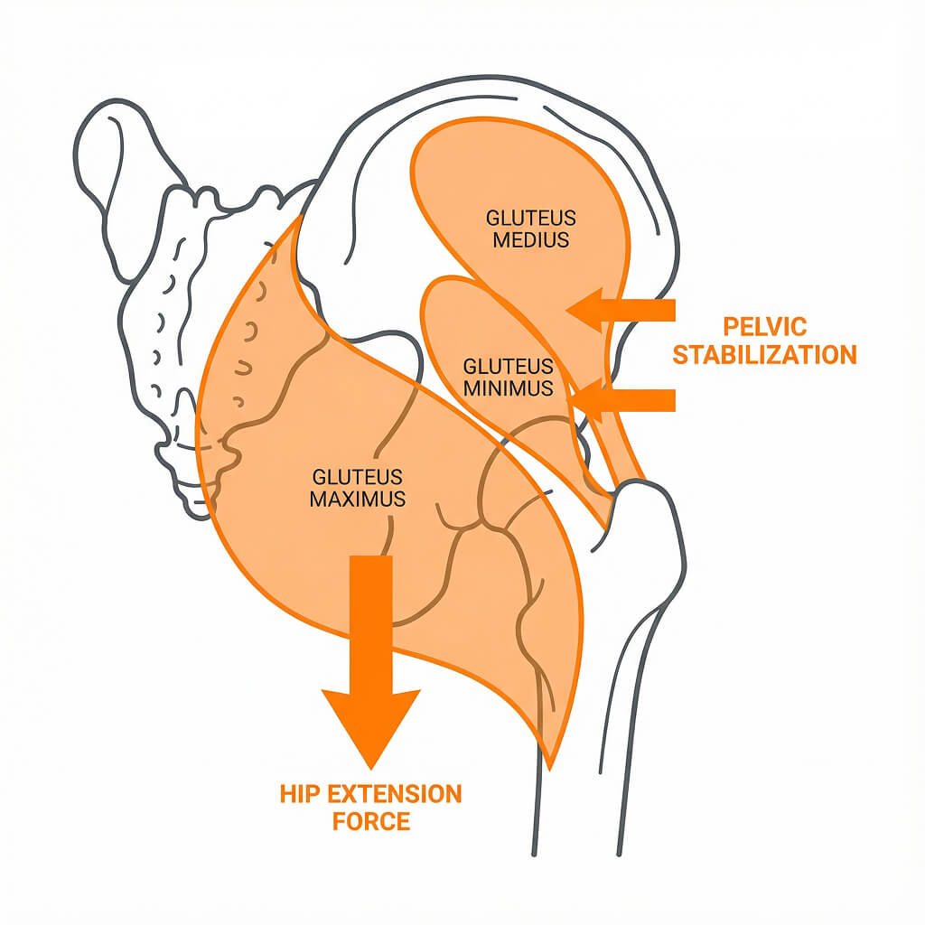
Primary Biological Actions
- Hip extension
- Hip abduction
- Hip external rotation
- Contribution to trunk control
Gluteus Medius
Origin, Insertion, and Structure
The gluteus medius lies above the gluteus maximus on the lateral surface of the pelvis.
Origin
- Outer surface of the ilium
Insertion
- Lateral surface of the greater trochanter
Structural Characteristics
- Fan-shaped muscle
- Mixed fiber orientation
- Positioned for control rather than power
Primary Biological Actions
- Hip abduction
- Anterior fibers: Hip internal rotation
- Posterior fibers: Hip external rotation
- Pelvic stabilization
- Control of femoral motion
Gluteus Minimus
Origin, Insertion, and Structure
The gluteus minimus is the smallest and deepest of the gluteal muscles.
Origin
- Outer surface of the ilium (inferior to medius)
Insertion
- Anterior surface of the greater trochanter
Structural Characteristics
- Thin muscle belly
- Close proximity to the hip joint
Primary Biological Actions
- Hip Abduction
- Hip stabilization
- Hip internal rotation
- Joint support during movement
Fiber Type Distribution in the Gluteal Muscles
Muscle fiber composition influences how a muscle responds to load and fatigue.
While individual variation exists, research suggests:
- Gluteus maximus contains a higher proportion of fast-twitch fibers
- Gluteus medius shows a more mixed fiber profile
- Gluteus minimus is more endurance-oriented
This distribution reflects the biological roles of each muscle rather than training preference.
The Role of Deep Hip Stabilizers
Beneath the gluteal muscles lies a group of smaller stabilizers often referred to as the deep six:
- Piriformis
- Obturator internus
- Obturator externus
- Superior gemellus
- Inferior gemellus
- Quadratus femoris
These muscles:
- Contribute primarily to external rotation and rotational control of the hip, with function varying depending on hip position.
- Support hip joint integrity
- Assist with rotational control
- Work synergistically with the glutes
They are anatomical contributors rather than primary movers.
Anatomy as Potential, Not Performance
Anatomy defines what is possible, not how movement is expressed under load.
Understanding:
- Where muscles are located
- How they attach
- What actions they perform
How these muscles interact during real movement depends on biomechanics, stability, and loading conditions.
From Anatomy to Functional Understanding
This reference guide establishes where the glute muscles are and what they do biologically.
To understand:
- How these muscles interact under load
- Why certain training methods succeed or fail
- How force production is influenced
Continue Learning
Gluteal Anatomy & Biomechanics
Hip Thrust Muscles Worked and Biomechanics
Anatomy names the parts.
Function explains what each part is built to do.